Foot Muscles Mri Anatomy / Https Encrypted Tbn0 Gstatic Com Images Q Tbn And9gcrut3tkhzhwgrstlwhngfr1rwlpocndbfjyybvquchem3l Ubfq Usqp Cau / It contributes to the surface anatomy of the medial sole of the foot and is easy to palpate.
byAdmin•
0
Foot Muscles Mri Anatomy / Https Encrypted Tbn0 Gstatic Com Images Q Tbn And9gcrut3tkhzhwgrstlwhngfr1rwlpocndbfjyybvquchem3l Ubfq Usqp Cau / It contributes to the surface anatomy of the medial sole of the foot and is easy to palpate.. The medial muscles of the foot sole have various tasks: The muscles lying within the medial group form a bulge referred to as the 'ball' of the big toe. Muscles of the foot (overview) the dorsal foot muscles are in the dorsum of foot and they extend the toes. Familiarity with the normal mr imaging anatomy of the nerves in the knee, leg, ankle, and foot is essential for accurate assessment of the presence of peripheral entrapment syndromes. Anatomy of the foot and ankle the foot is a structure of the body with numerous joints, bones, muscles, ligaments, and tendons.
It is a complex anatomical structure and can be subdivided into the hindfoot, the midfoot, and the forefoot. The muscles lying within the medial group form a bulge referred to as the 'ball' of the big toe. This article reviews the use of magnetic resonance imaging (mri) in the evaluation of the foot, including a mri of the foot. The muscles lying within the medial group form a bulge. Bone contusions, osteonecrosis, marrow oedema syndromes, and stress > fractures) > synovial based disorders ( e.g.
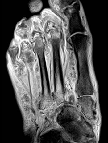
Radiological Anatomy X Ray Ct Mri Kenhub from thumbor.kenhub.com Foot muscles mri anatomy : 12 photos of the foot muscle anatomy mri. As the fiber bundles extend distally, they become grouped into four bellies. A magnetic resonance imaging (mri) was performed on a normal subject; Related posts of foot muscle anatomy mri. It is responsible for the coordinated movements of gait and the body's ability to stand upright(1). Top suggestions for foot muscle anatomy mri. The bones of the foot are:
Bone contusions, osteonecrosis, marrow oedema syndromes, and stress > fractures) > synovial based disorders ( e.g.
The lumbrical muscles of the foot are four muscles that originate from the tendons of the flexor digitorum longus and pass dorsally to insert into the free medial margins of the extensor hoods of the four lateral toes. Here we explain the major muscles of the human body. The extrinsic muscles arise from the anterior, posterior and lateral compartments of the leg. Top suggestions for foot muscle anatomy mri. Familiarity with the normal mr imaging anatomy of the nerves in the knee, leg, ankle, and foot is essential for accurate assessment of the presence of peripheral entrapment syndromes. The muscles lying within the medial group form a bulge. They are mainly responsible for actions such as eversion, inversion, plantarflexion and dorsiflexion of the foot. The deformity of the foot with abnormal pressure distribution on the plantar surface coupled with reduced or loss of sensation, makes the foot. 12 photos of the foot muscle anatomy mri. Dr calum worsley and dr daniel macmanus et al. Those fibers of the most medial and largest belly are known as. They act collectively to stabilise the arches of the foot, and individually to control movement of the digits. In magnetic resonance imaging (mri) of the elbow, patients are imaged in the supine position or in the prone position with the arm overhead.
This anatomy, and stabilization by the lisfranc ligament, is important for support of the arch of the foot. With a good grasp of foot anatomy it readily becomes apparent which surgical approaches can be used to access various areas of the foot and ankle. As the fiber bundles extend distally, they become grouped into four bellies. Coronal images are perpendicular to the long axis of the metatarsals. The muscles of the dorsum of the foot are a group of two muscles, which together represent the dorsal foot musculature.
Radiological Anatomy X Ray Ct Mri Kenhub from thumbor.kenhub.com The medial muscles of the foot sole have various tasks: The muscles of the dorsum of the foot are a group of two muscles, which together represent the dorsal foot musculature. There are only two muscles in the dorsal group, while the plantar muscles are further subdivided into three groups; Neuropathies around the elbow joint. With a good grasp of foot anatomy it readily becomes apparent which surgical approaches can be used to access various areas of the foot and ankle. Plantar foot muscles mri / central plantar muscles of the foot: Dr calum worsley and dr daniel macmanus et al. It is responsible for the coordinated movements of gait and the body's ability to stand upright(1).
Related posts of foot muscle anatomy mri.
Synovitis, tenosynovitis, bursitis, and ganglion cysts) > congenital and developmental conditions( eg.dysplasia, tarsal coalition). Dr calum worsley and dr daniel macmanus et al. Magnetic resonance imaging (mri) diagnostic ultrasonography (us) nerve conduction study and other bone scans as necessary more aggressive one of the biggest contributors to plantar fasciitis is weakened foot muscles and a disconnect from. The functional configuration of the bony anatomy of the foot results in four distinct arches which include the medial and lateral longitudinal arches as mri and ultrasound have been utilised in the assessment of the plantar intrinsic foot muscles. A magnetic resonance imaging (mri) was performed on a normal subject; The foot is the most distal part of the lower limb below the leg and ankle. A magnetic resonance imaging (mri) was performed on a normal subject; It contributes to the surface anatomy of the medial sole of the foot and is easy to palpate. The anatomy of the nerves of the foot and ankle is quite complex. The muscles lying within the medial group form a bulge. Routine ankle magnetic resonance imaging (mri) tests involve taking images of the foot and ankle in the axial, coronal, and sagittal planes parallel to the tabletop(2). Subscribe to foot & ankle problems. Neuropathies around the elbow joint.
12 photos of the foot muscle anatomy mri. Synovitis, tenosynovitis, bursitis, and ganglion cysts) > congenital and developmental conditions( eg.dysplasia, tarsal coalition). It is responsible for the coordinated movements of gait and the body's ability to stand upright(1). Here we explain the major muscles of the human body. There are a variety of anatomical structures that make up the anatomy of the foot and ankle (figure 1) including bones, joints, ligaments, muscles, tendons, and nerves.
Mri Of The Diabetic Foot Radsource from radsource.us Plantar foot muscles mri / central plantar muscles of the foot: Muscles of the foot are located on its rear and on the sole. Bone contusions, osteonecrosis, marrow oedema syndromes, and stress > fractures) > synovial based disorders ( e.g. It results in pain in the heel and bottom of the foot that is usually most severe with the first steps of the day or following a period of rest. A magnetic resonance imaging (mri) was performed on a normal subject; Lateral and medial processes of calcaneal tuberosity. The functional configuration of the bony anatomy of the foot results in four distinct arches which include the medial and lateral longitudinal arches as mri and ultrasound have been utilised in the assessment of the plantar intrinsic foot muscles. Coronal images are perpendicular to the long axis of the metatarsals.
They act collectively to stabilise the arches of the foot, and individually to control movement of the digits.
Subscribe to foot & ankle problems. 12 photos of the foot muscle anatomy mri. The muscles lying within the medial group form a bulge referred to as the 'ball' of the big toe. Related posts of foot muscle anatomy mri. Lumbrical muscles of the foot dr waleed bukhari and dr geon oh et al. The muscles lying within the medial group form a bulge. With a good grasp of foot anatomy it readily becomes apparent which surgical approaches can be used to access various areas of the foot and ankle. The muscles of the dorsum of the foot are a group of two muscles, which together represent the dorsal foot musculature. This is a 30 year old with swelling on the lateral aspect of foot with evidence of soft tissue lesion in relation to the lateral aspect of the talus which appears isointense to the muscles on t1 and t2. Extensor brevis and longus muscles. There are only two muscles in the dorsal group, while the plantar muscles are further subdivided into three groups; Related posts of foot muscle anatomy mri. Neuropathies around the elbow joint.
Related posts of foot muscle anatomy mri muscle anatomy books free download foot muscles mri. Most superficial of all the layers.